Magnetic Resonance - Technology Information Portal Welcome to MRI Technology
Info
  Sheets

Out-
      side


 'Surface Coil' 
SEARCH FOR    
 
  2 3 5 A B C D E F G H I J K L M N O P Q R S T U V W X Y Z
Result : Searchterm 'Surface Coil' found in 2 terms [] and 87 definitions []
previous     6 - 10 (of 89)     next
Result Pages : [1]  [2 3 4 5 6 7 8 9 10 11 12 13 14 15 16 17 18]
Searchterm 'Surface Coil' was also found in the following services: 
spacer
News  (1)  Resources  (2)  Forum  (4)  
 
Phased Array CoilInfoSheet: - Coils - 
Intro, 
Overview, 
etc.MRI Resource Directory:
 - Coils -
 
The phased array coils operate typically as receive only coils. In that case, the in the MRI device implemented body coil act as the transmitter and sends the radio frequency energy to generate the excitation pulses. State-of-the-art array coil systems include the use of 4 (up to 32) coils with separate receivers. This method is often referred to as a phased array system, although the signals are not added such that the signal phase information is included. The use of phased array coils allows the decreasing of the number of signal averages, which shortens the scan time by high SNR and resolution.
High-sensitivity RF surface coils and digital processing algorithms have been developed that speed up image acquisition and reconstruction during the MRI scan.
Fast parallel imaging techniques, for example sensitivity encoding (SENSE), 'Partially Parallel Imaging with Localized Sensitivity' (PILS), Simultaneous Acquisition of Spatial Harmonics (SMASH) or Array Spatial Sensitivity Encoding Technique (ASSET) use phased array multichannel coils to further improve spatial and temporal resolution. The sensitivity profile of a phased array coil element is measured by a separate low resolution 3D acquisition over the entire field of view in the case of a SENSE acquisition. For an mSENSE measurement, a self-calibration acquires some of the missing lines in the center of the k-space.
Also called linear array coil or synergy surface coil.

See also the related poll result: '3rd party coils are better than the original manufacturer coils'
spacer
 
• Related Searches:
    • Switchable Coil
    • Coil Diameter
    • Array Coil
    • Imaging Coil
    • Sensitivity Encoding
 
Further Reading:
  Basics:
Coil Arrays for Parallel MRI: Introduction and Overview.
   by www.mr.ethz.ch    
MRI Resources 
Mass Spectrometry - Blood Flow Imaging - Shoulder MRI - Safety Training - Brain MRI - Research Labs
 
Signa HDx 1.5T™InfoSheet: - Devices -
Intro, 
Types of Magnets, 
Overview, 
etc.
 
www.vitalcom.com/euen/mri/products/signa-hdx-15t/index.html From GE Healthcare;
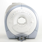
The GE Signa HDx MRI system is a whole body magnetic resonance scanner designed to support high resolution, high signal to noise ratio, and short scan times.
The 1.5T Signa HDx MR Systems is a modification of the currently marketed GE 1.5T machines, with the main difference being the change to the receive chain architecture that includes a thirty two independent receive channels, and allows for future expansion in 16 channel increments. The overall system has been improved with a simplified user interface and a single 23" liquid crystal display, improved multi channel surface coil connectivity, and an improved image reconstruction architecture known as the Volume Recon Engine (VRE).
Device Information and Specification
CLINICAL APPLICATION
Whole body
CONFIGURATION
Compact short bore
Possible
SYNCHRONIZATION
ECG/peripheral, respiratory gating, (SmartPrep, SmartStep)
PULSE SEQUENCES
Standard: SE, IR, 2D/3D GRE and SPGR, Angiography: 2D/3D TOF, 2D/3D Phase Contrast; 2D/3D FSE, 2D/3D FGRE and FSPGR, SSFP, FLAIR, EPI, optional: 2D/3D Fiesta, FGRET, Spiral, Tensor,
IMAGING MODES
2D single slice, multi slice, and 3D volume images, multi slab, cine
1 cm to 48 cm continuous
2D 0.7 mm to 20 mm; 3D 0.1 mm to 5 mm
1028 x 1024
MEASURING MATRIX
128x512 steps 32 phase encode
PIXEL INTENSITY
256 gray levels
POWER REQUIREMENTS
480 or 380/415
COOLING SYSTEM TYPE
Closed-loop water-cooled gradient
CRYOGEN USE, L/hr
less than 0.03 L/hr liquid helium
spacer
MRI Resources 
Blood Flow Imaging - MRI Physics - - MRI Technician and Technologist Jobs - Breast MRI - Manufacturers
 
Single Turn SolenoidInfoSheet: - Coils - 
Intro, 
Overview, 
etc.MRI Resource Directory:
 - Coils -
 
Single turn solenoid is a transmit and receive RF imaging coil that, in general, has a cylindrical shape. The solenoidal configuration of this coil is a further developed planar surface coil. While surface coils have a strong coupling and high signal to noise ratio to nearby signals they have a marked loss of signal beyond one radius from the center of the coil. With a solenoidal coil design the imaging object lies within one radius from the coil center, which improved imaging (of extremities, such as wrist or knee).

See also Radio Frequency Coil and Imaging of the Extremities.
spacer

• View the DATABASE results for 'Single Turn Solenoid' (3).Open this link in a new window

Searchterm 'Surface Coil' was also found in the following services: 
spacer
News  (1)  Resources  (2)  Forum  (4)  
 
Spatially Localized Spectroscopy
 
Process by which regions of tissue are selectively sampled to produce spectra from defined volumes in space. These methods may be employed to sample a single region in space (single voxel method) or multiple regions simultaneously (multivoxel methods). The spatial selectivity can be achieved by a variety of methods including surface coils, surface coils in conjunction with RF gradient methods, or RF pulses in combination with switched magnetic field gradients, for example, volume-selective excitation. An indirect method of achieving spatial selectivity is the destruction of coherence of the magnetization in regions that lie outside the region of interest. A variety of spatial encoding schemes have been employed for multivoxel localization. See Chemical shift imaging.
spacer
MRI Resources 
Diffusion Weighted Imaging - Portals - Cochlear Implant - Equipment - General - Case Studies
 
Zero Fill ArtifactInfoSheet: - Artifacts - 
Case Studies, 
Reduction Index, 
etc.MRI Resource Directory:
 - Artifacts -
 
Quick Overview
Artifact Information
NAME
Zero fill
DESCRIPTION
Zebra stripes or other anomalies
REASON
Signal changes
HELP
Surface coil, change the sequence
Missing or zero filled data in k-space can cause artifacts from anomalies to zebra stripes.
mri safety guidance
Image Guidance
Change the sequence parameters or try to use a surface coil. If the problem persists, it must be addressed by a service representative.
spacer

• View the DATABASE results for 'Zero Fill Artifact' (2).Open this link in a new window

 
Further Reading:
  Basics:
Zero-Fill Artifacts
   by www.mritutor.org    
MRI Resources 
Service and Support - Distributors - Blood Flow Imaging - Universities - PACS - Spectroscopy
 
previous      6 - 10 (of 89)     next
Result Pages : [1]  [2 3 4 5 6 7 8 9 10 11 12 13 14 15 16 17 18]
 Random Page
 
Share This Page
FacebookTwitterLinkedIn

MR-TIP    
Community   
User
Pass
Forgot your UserID/Password ?    


New acceleration techniques will :
reduce scan times 
cause artifacts 
increase expenses 
be useful if you have a lot of experience 
doesn't do much 
never heard of 

Look
      Ups


MR-TIP.com uses cookies! By browsing MR-TIP.com, you agree to our use of cookies.

Magnetic Resonance - Technology Information Portal
Member of SoftWays' Medical Imaging Group - MR-TIP • Radiology-TIP • Medical-Ultrasound-Imaging • 
Copyright © 2003 - 2024 SoftWays. All rights reserved. [ 5 May 2024]
Terms of Use | Privacy Policy | Advertising
 [last update: 2024-02-26 03:41:00]